Do our brains process natural and deepfaked voices differently? In a new study published in Communications Biology, scientists from Switzerland and Australia have shown that this is the case. Neuroimaging experiments identified two brain regions that respond differently to natural and deepfake voices. The results add to our understanding of neural mechanisms behind our ability to recognise deceptive information in digital environments. To precisely characterise the brain network, the researchers used probabilistic maps from the openly accessible Julich Brain Atlas on EBRAINS.
News
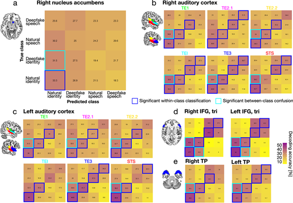
Julich Brain Atlas used in study to understand how the brain detects deepfakes
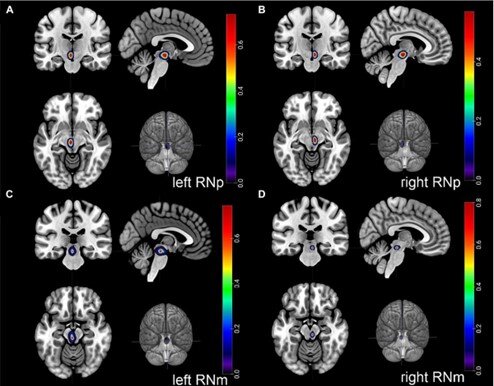
New study shows brain maps of the "red nucleus"
In a recent study, a German-American team of scientists has now revealed new insights into the evolutionary development of a brain structure that plays a crucial role in finely tuned and skillful hand movements. The researchers from C.&O. Vogt Institute for Brain Research at HHU Düsseldorf, Forschungszentrum Jülich and Stony Brook University in New York studied a brain structure called the “red nucleus” (lat. Nucleus ruber). This nucleus is an important element of our motor system, especially for dexterous hand movements.
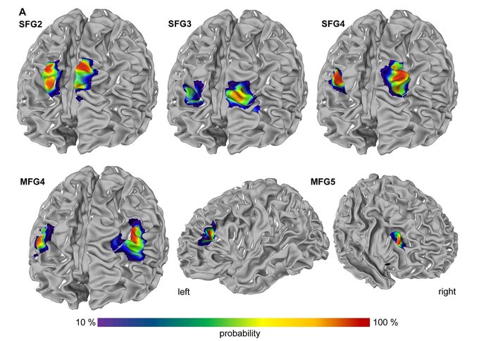
New brain maps on EBRAINS show organizational principles of the prefrontal cortex
Newly published new brain maps identify five new areas within the dorsolateral prefrontal cortex (DLPFC), that plays a crucial role in executive functions, including working memory, decision-making, and attention. The new study introduces 3D maps of cytoarchitectonic regions, enhancing our understanding of the prefrontal cortex's complexity.

EBRAINS Research Infrastructure Secures €38 Million in Funding for New Phase of Digital Neuroscience
The European Commission has signed a grant agreement to fund EBRAINS with €38 million until 2026. Over the next three years, the infrastructure will continue to develop tools and services to widely serve research communities in neurosciences, brain medicine, and brain-inspired technologies.
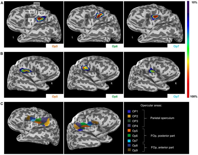
HBP researchers identify three new human brain areas involved in sexual sensation, motor coordination, and music processing
HBP researchers from Germany performed detailed cytoarchitectonic mapping of distinct areas in a human cortical region called frontal operculum and, using connectivity modelling, linked the areas to a variety of different functions including sexual sensation, muscle coordination as well as music and language processing.
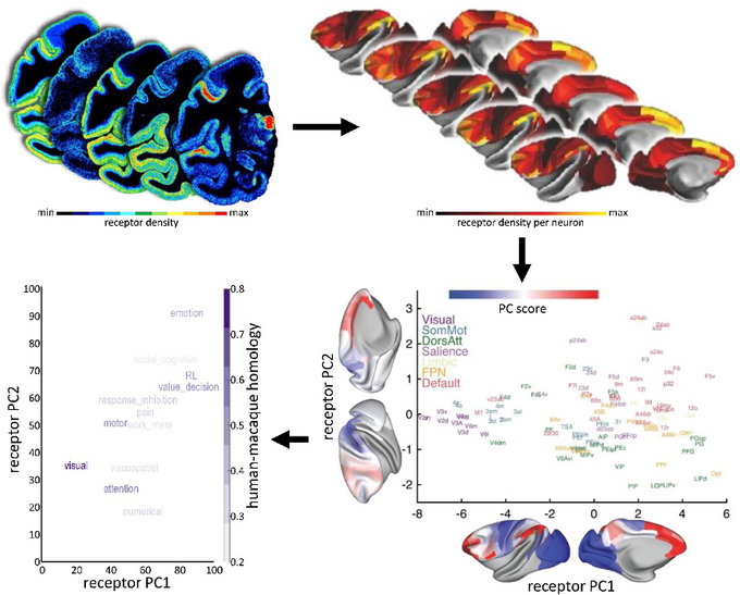
Human Brain Project study offers insights into neurotransmitter receptor organisation
Julich Brain Atlas researchers in collaboration with teams from the UK, the US and France have made advances on our understanding of the distribution of neurotransmitter receptors across the brain.
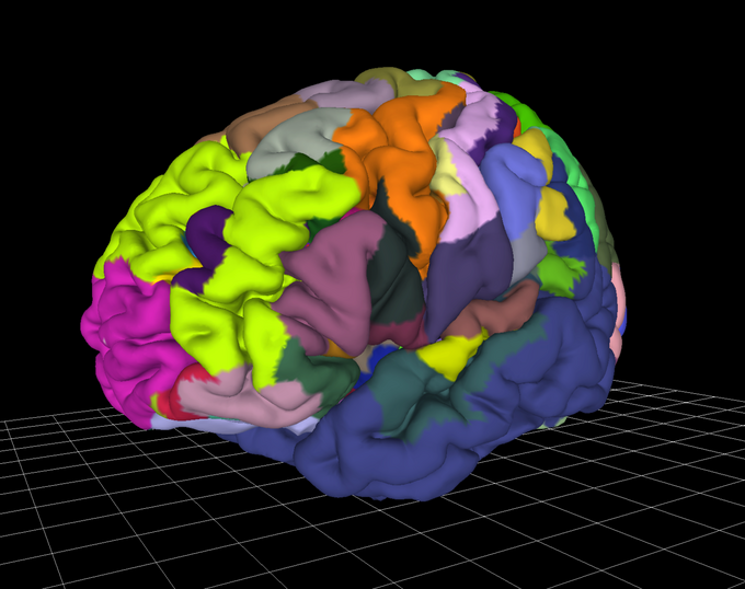
Multilevel brain atlases provide tools for better diagnosis
The multilevel Julich Brain Atlas developed by researchers in the Human Brain Project, could help in studying psychiatric and aging disorders by correlating brain networks with their underlying anatomical structure. By mapping microarchitecture with unprecedented levels of detail, the atlas allows for better understanding of brain connectivity and function. Researchers of the HBP have provided an overview of the Julich Brain Atlas in the journal Biological Psychiatry. The paper focuses on the cytoarchitecture and receptor architecture of the human brain, and how to apply the atlas in the field of psychiatric research.
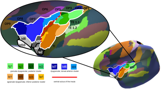
Seven new areas in the human insular cortex mapped for the first time
A team of researchers from the C. and O. Vogt Institute for Brain Research at the University of Düsseldorf and the Institute of Neuroscience and Medicine (INM-1) at Forschungszentrum Jülich have identified seven new areas of the human insular cortex, a region of the brain that is involved in a wide variety of functions, including self-awareness, cognition, motor control, sensory and emotional processing. All newly detected areas are now available as 3D probability maps in the Julich Brain Atlas, and can be openly accessed via the Human Brain Project’s EBRAINS infrastructure. Their findings, published in NeuroImage, provide new insights into the structural organisation of this complex and multifunctional region of the human neocortex.

Alexey Chervonnyy receives PhD Scholarship from Hector Fellow Academy
Alexey Chervonnyy has been awarded a PhD Scholarship from the Hector Fellow Academy. The scholarship, awarded after a multi-stage selection process, will fund a three-year research project at the C. and O. Vogt Institute for Brain Research in Düsseldorf under the supervision of Hector Fellow Katrin Amunts. Chervonnyy will investigate the structure of the human hypothalamus, a part of the brain that plays a key role in regulating neuroendocrine, behavioural and autonomic processes essential to life, such as circadian rhythms, metabolic processes, sleep and body temperature.