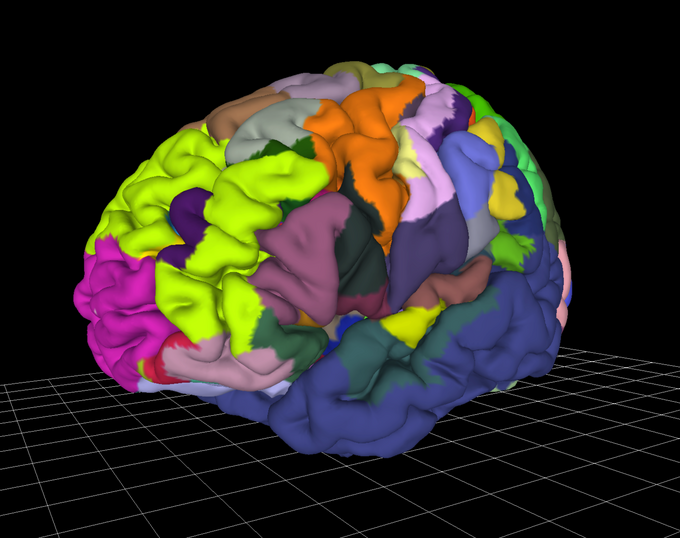
Release 3.1 of the Julich Brain Atlas has been published and can be freely downloaded through the EBRAINS research infrastructure. The updated brain atlas now gives online access to 52 new probability maps of cortical and subcortical structures in a three-dimensional reference space.
News

New release of the Julich-Brain Atlas adds 52 new maps
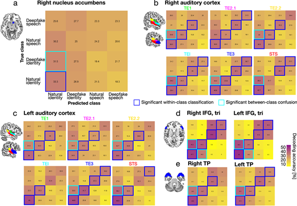
Julich Brain Atlas used in study to understand how the brain detects deepfakes
Do our brains process natural and deepfaked voices differently? In a new study published in Communications Biology, scientists from Switzerland and Australia have shown that this is the case. Neuroimaging experiments identified two brain regions that respond differently to natural and deepfake voices. The results add to our understanding of neural mechanisms behind our ability to recognise deceptive information in digital environments. To precisely characterise the brain network, the researchers used probabilistic maps from the openly accessible Julich Brain Atlas on EBRAINS.
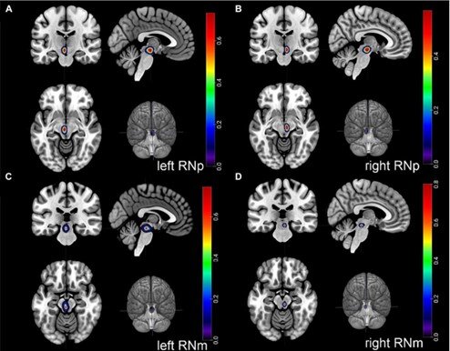
New study shows brain maps of the "red nucleus"
In a recent study, a German-American team of scientists has now revealed new insights into the evolutionary development of a brain structure that plays a crucial role in finely tuned and skillful hand movements. The researchers from C.&O. Vogt Institute for Brain Research at HHU Düsseldorf, Forschungszentrum Jülich and Stony Brook University in New York studied a brain structure called the “red nucleus” (lat. Nucleus ruber). This nucleus is an important element of our motor system, especially for dexterous hand movements.
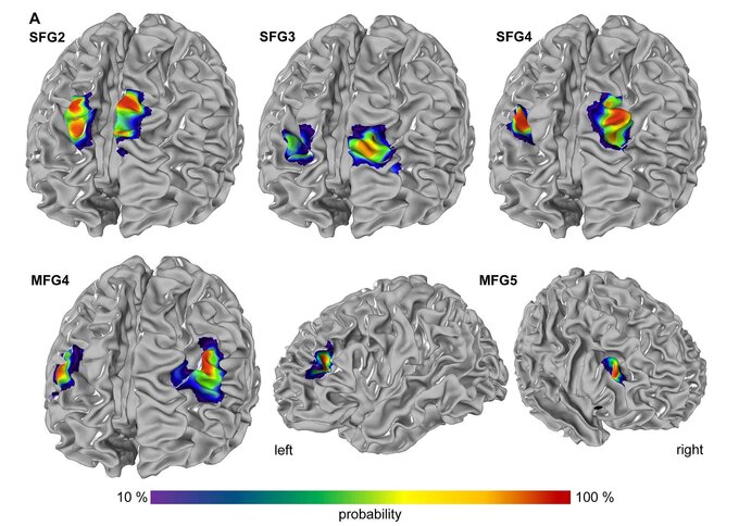
New brain maps on EBRAINS show organizational principles of the prefrontal cortex
Newly published new brain maps identify five new areas within the dorsolateral prefrontal cortex (DLPFC), that plays a crucial role in executive functions, including working memory, decision-making, and attention. The new study introduces 3D maps of cytoarchitectonic regions, enhancing our understanding of the prefrontal cortex's complexity.

EBRAINS Research Infrastructure Secures €38 Million in Funding for New Phase of Digital Neuroscience
The European Commission has signed a grant agreement to fund EBRAINS with €38 million until 2026. Over the next three years, the infrastructure will continue to develop tools and services to widely serve research communities in neurosciences, brain medicine, and brain-inspired technologies.
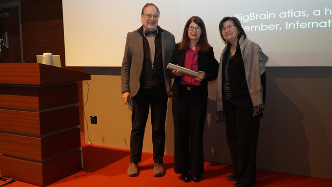
INM-1 director Katrin Amunts receives Justine and Yves Sergent International Award
Katrin Amunts has been honoured with the 2023 Justine and Yves Sergent International Award, which is granted to outstanding female researchers for contributions in the field of cognitive neuroscience and brain imaging who have achieved a high international reputation. The award was presented during a conference at the University of Montreal, Canada, on Friday, December 8th, where Katrin Amunts gave the keynote “The human brain atlas – mapping the brain as a way to understand its function.”
“Impressive research results” - external review panel evaluates final results of Human Brain Project
From 21-24 November, the final Human Brain Project (HBP) review was held in Brussels during which members of the HBP consortium presented the final project results to a panel of external scientific experts. The scope of this review was the final phase of the HBP, which ended in September 2023. Results were presented in detailed documentation and presentations and followed by extensive Q&As.
7th BigBrain Workshop 2023
Registration is now open for the 7th BigBrain Workshop, taking place in the beautiful city of Reykjavik, Iceland, on October 4th to 6th, 2023. This workshop is an opportunity for the neuroscientific community to come together and present their cutting-edge research, discuss future prospects of the BigBrain data and tools, and explore how to better leverage high-performance computing and artificial intelligence to create multimodal, multiresolution tools for the high-resolution BigBrain and related datasets.
We are proud of our confirmed keynote speakers Kári Stefánsson, Paul Thompson, and Maryann Martone.
We are also pleased to announce that this year's BigBrain Workshop will be held in conjunction with an HBHL Training Day, taking place as a full day event on October 4, on-site at the conference venue.
The event is free of charge but prior registration is required.
Information about the programme will be updated regularly on the website:https://go.fzj.de/BigBrainWorkshop2023

The Human Brain Project ends: What has been achieved
On September 30th, the Human Brain Project (HBP) formally completes its 10-year runtime as an EU-funded FET Flagship. The project has pioneered digital neuroscience, a new approach to studying the brain based on multidisciplinary collaborations and high-performance computing. The HBP will continue to have an impact on neuroscience for many years through the EBRAINS research infrastructure and a new way of collaborative work in the field.